In this post, we will discuss subjects such as WBC Types, Leukocyte Origin and Development, Different WBC Concentrations in Blood, WBC Function, and WBC Lifespan.
White blood cells[WBC] are also known as leukocytes. These are the body’s immune cells.
The body’s defensive system is made up of mobile units called leukocytes.
- Types of White Blood cells or Leukocytes.
- Origin & Development of WBC’s.
- Different white blood cell[WBC’s] concentrations in the blood.
- Granulocytes.

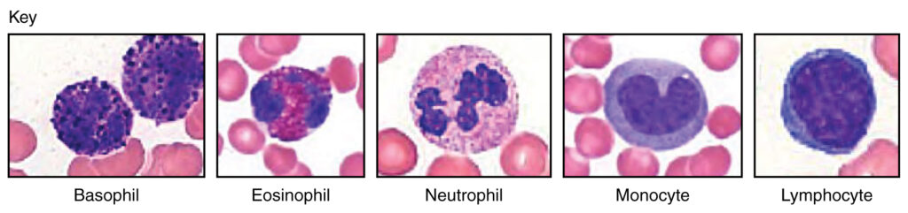
Types of White Blood cells or Leukocytes.
White blood cells, or WBC, are divided into two types: granulocytes and agranulocytes.
Granulocytes consist of Eosinophils, basophils & neutrophils.
Agranulocytes consist of Lymphocytes & Monocytes
Origin & Development of WBC’s.
They partially develop in Primary lymphoid Organs like Thymus gland , Bone marrow & Partially in Lymphoid tissues.
It is mostly present in lymph nodes, although it may also be found in lymphoid follicles in tonsils, Peyer’s patches, spleen, adenoids, skin, and other locations linked with mucosa-associated lymphoid tissue (MALT).
Different white blood cell[WBC’s] concentrations in the blood.
An adult human’s blood has 7000 WBC per microliter.
| Cell types | % of total in Blood leukocytes |
| Neutrophils | 62.0% |
| Eosinophil | 2.3% |
| Basophils | 0.4% |
| Monocytes | 5.3% |
| Lymphocytes | 30% |
Granulocytes.
Eosinophils

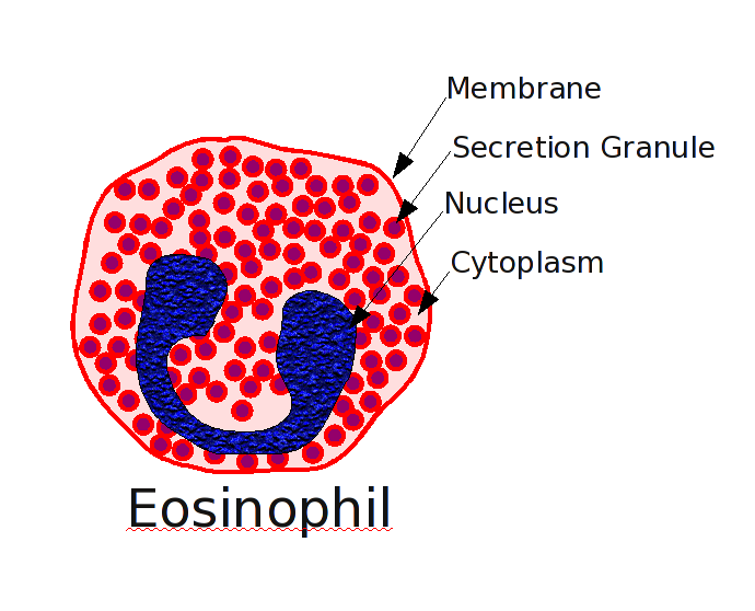
Eosinophils are granular leukocytes from the myeloid cell line within bone marrow.
Eosinophils generally account for roughly 2% of total blood leukocytes.
They have bilobed nuclei ,sausage-shaped nucleus and measure 12-17 micrometres in size.
EOSINOPHILIA
People with parasite illnesses such as Schistosomiasis and Trichinosis produce quite a lot of eosinophils.
Functions.
Additionally, eosinophils are involved in inflammatory responses, tissue damage, and allergic reactions, including asthma, rhinitis, and allergies.
Chemotaxis movement is seen by eosinophils.
Interaction with T lymphocytes in allergies and Helminth larvicidal infections including schistosomiasis and trichinosis.
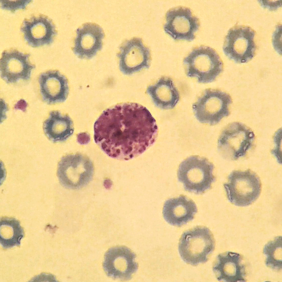
Basophils.
Basophils are granular leukocytes from the myeloid cell line within bone marrow. In general, they constitute less than 0.5 percent of all blood leukocytes. Basophils are 12 to 15 m in diameter, with bi-lobed or S-shaped nuclei, and contain cytoplasmic special granules (0.5 m in diameter) that are blue to purple in color.
A combination of sulfated glycosaminoglycans and heparin is responsible for the granules’ basophilia.
What are the causes of elevated basophil levels in blood?
Higher amounts of basophils have been associated to specific illnesses or basophilic disorders.
(Increased count) > 50/mm3 or > 0.05 × 10⁹/L
- Chickenpox.
- Hodgkin disease
- acute basophilic leukemia
- Allergic reaction
- Bone marrow diseases
- Chronic myelogenous leukemia, a kind of bone marrow cancer.
What are the causes of low level of basophil levels in blood?
Basopenia is characterized by a reduction or low level of basophils in the blood.
- Hyperthyroidism
- Thryrotoxicosis
- Acute Infection.
- stress reactions
- Myocardial Infarction
- prolonged steroid therapy, chemotherapy, radiation

Neutrophils.

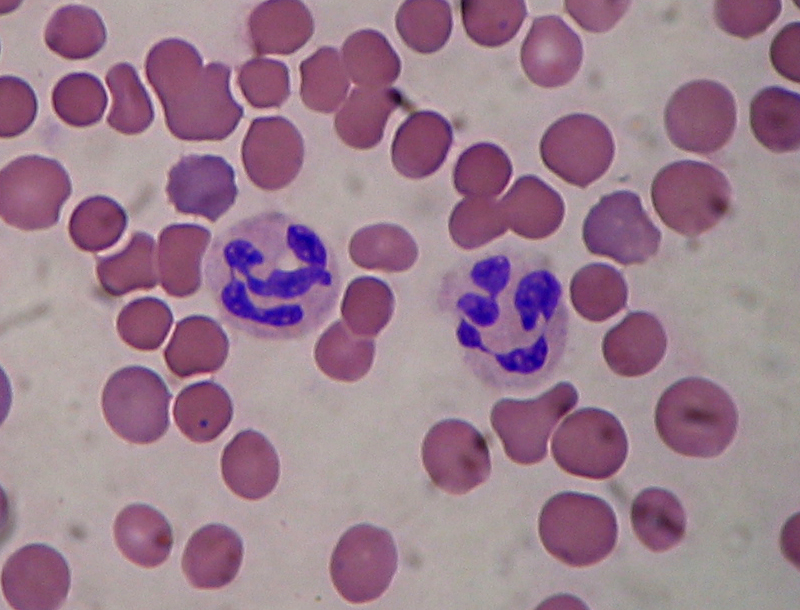
Neutrophil are granular leukocytes from the myeloid cell line within bone marrow. In general they constitute 50-70% of all blood leukocytes.
They contain a nucleus divided into 3-5 lobules OR MULIT LOBED
Neutrophils further divided into banded neutrophil & segmented neutrophil of the Polymorphonuclear neutrophils
Functions
Diapedesis is the ability of neutrophils and monocytes to squeeze through gaps between endothelial cells in blood capillaries.
The process of chemotaxis draws neutrophils to areas of inflammatory tissue.
A major function of the neutrophil and macrophages is phagocytosis which is cellular ingestion of cell debris & a foreign body.
key role of neutrophils
- Chemotaxis
- Anti microbial functions
- phagocytosis
- Degranulation
Leave a Reply Cancel reply