This article will cover the stages of erythropoiesis, the genesis of red blood cells[RBC’s] erythropoiesis regulation factors, and the clinical significance of reticulocytes.
By use of the erythropoiesis process, the human body produces two million red blood cells[RBC’s] per second.


Genesis of red blood cell [Erythrocytes].
ERYTHROPOESIS is described as the process of forming red blood cells aka RBCs or Erythrocytes.
Where does it occur?
In Early Embryonic life
YOLK SAC(3–8 weeks)
- Liver(6 weeks–birth)
- Spleen(10–28 weeks)
- Red bone marrow (Spongy bone trabeculae) 18 weeks to Adult
- Skull
- Sternum
- Pelvis
- Epiphyses of long bone.
A mnemonic to remember the site of erythropoiesis.
Young Liver Synthesizes Blood cell.
Young –Yolk Sac.
Liver-Liver.
Synthesizes-Spleen.
Blood-Bone Marrow.
Erythropoiesis refers to the process of producing red blood cells from pluripotent hemopoietic stem cells (PHSC).

A tiny number of pluripotent stem cells [PHSC] remain exactly as this PHSC and the rest transform into committed stem cells.
When these cells are cultivated in culture, they create colonies of specific types of blood cells, such as CFU-E, CFU-GM, and CFU-M.
The term “colony forming unit erythrocyte,” or CFU-E, refers to committed stem cells that will eventually produce erythrocytes.
What Is the Cause of Erythropoiesis?
Low oxygen availability to the tissue or a hypoxic environment
Factors that reduce the availability of oxygen to the tissues.
Low blood volume
Anemia
Low hemoglobin
Pulmonary disease.
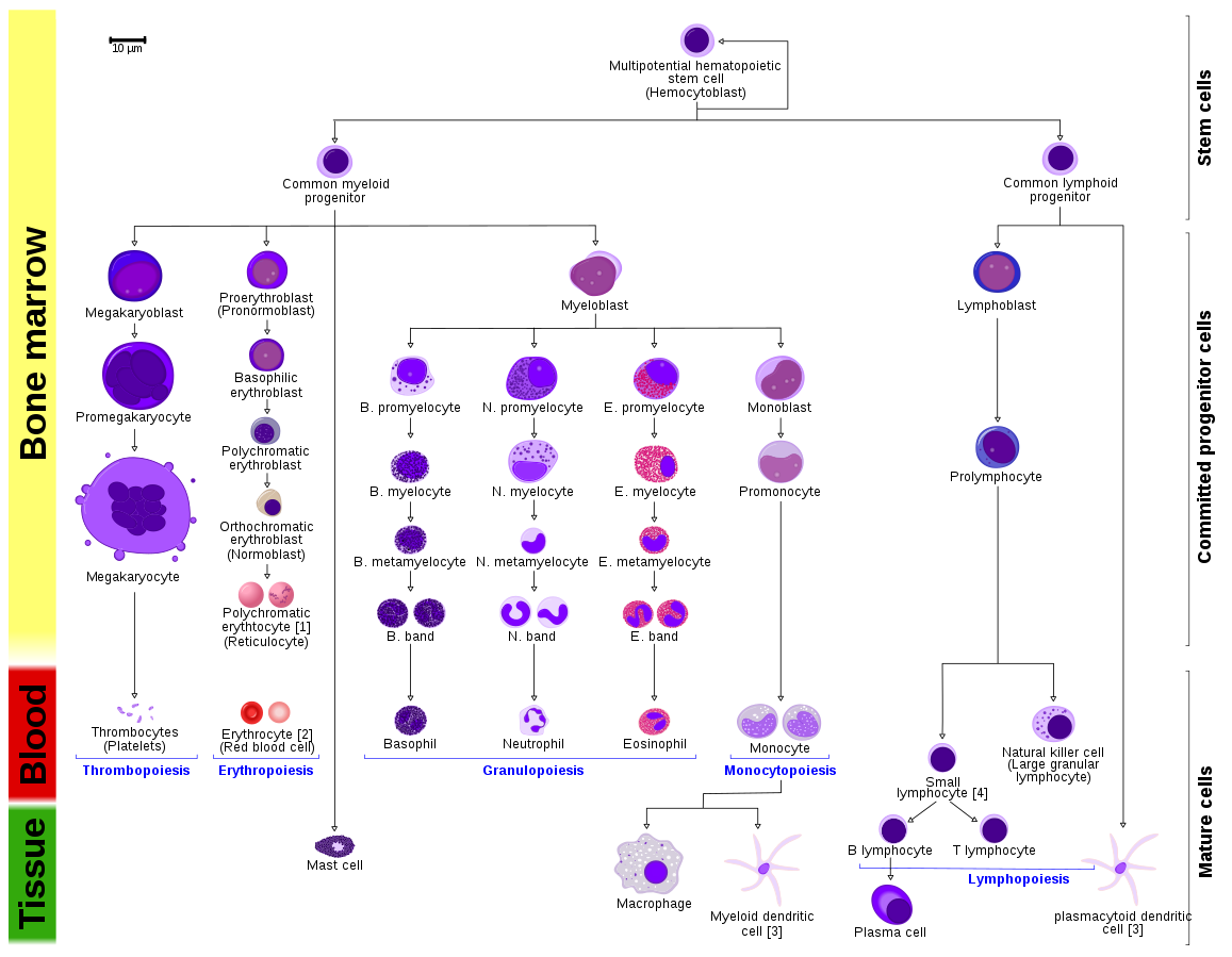
The stages of differentiation of erythrocytes.
Under the influence of hormones such as erythropoietin, androgens, thyroid hormones, and corticosteroids, CFU-E transforms the proerythroblast cell, which is known as the first cell in the RBC lineage.
Proerythroblast.
Proerythroblast cells proliferate and divide into Basophilic erythroblast.
Basic dyes are used to stain these cells.
Polychromatophil erythroblast.
Hemoglobin first appears in polychromatophil erythroblast or intermediate normoblasts.
In the succeeding generation
The nucleus condenses to a small size or disappears from the cell.
There are trace amounts of cell organelles like mitochondria and golgi bodies within the cell.
This cell stage is also referred to as Reticuolcyte.
During the reticulocyte stage, cells move from the bone marrow into blood capillaries via diapedsis.
Erythropoiesis regulation factors.
- Hypoxia & erythropoietin.
- VItaminB12.
- Folic acid.
- Iron.
- Pyrodoxine.
- Dietary protein and energy.
the clinical significance of reticulocytes.
Reticulocyte count in a healthy person should be between 0.5 to 2.5%
Reduced reticulocyte counts in patients are linked to nutritional deficiencies, hypochromic anemias, myelodysplastic syndromes, and aplastic anemias.
An increase in reticulocyte count is linked to the following conditions: hemolytic anemias and blood loss.
ATTRIBUTIONS.
Image by Freepik.
Attribution:
Original: A. Rad Vector: Rexxs, birdy and Mikael Häggström, M.D. Author info – Reusing images- Conflicts of interest: None Mikael Häggström, M.D., CC BY-SA 3.0, via Wikimedia Commons.
Leave a Reply Cancel reply